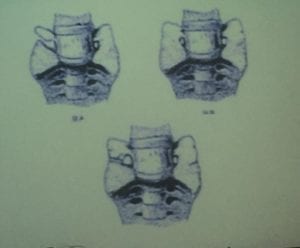
Studies show up to one-third of individuals have a transitional lumbosacral joint i.e. either the S1 is lumbarised ie. extra free standing disc between what would be S1 and S2 (in this case the S1 vertebra may be referred to as L6). Or the L5 is sacralised i.e. joined to sacrum via connection of the TPs to the sacral alar, this may be involve these processes just touching (type 1), forming a pseudo-arthrosis (type 2 aka Bertolottis's syndrome)), or completely fused (type 3 - Castellvi's classification) on one side of both (if fused on one side and pseudoarthrosis on the other this a type 4). The consequences of this are
A: Sacralisation of the L5/S1 ALWAYS causes movement restriction and protection of the L5S1 disc (i.e. it does not degenerate) BUT compensation of the L4/5 disc which tends to generate much quicker as too much movement occurs here (posses the point that movement is not good for discs and we be careful not to over mobilise!).
B: In the case of pseudo-arthrosis (types 2 and 4) this linked to increased likelihood and severity of low back pain due to OA changes of the pseudoarthrosis (as well as requiring and responding to regular manual therapy, this will likely require referral for CT / image-guided injections, radiofrequency denneration).
C: Patients with transitional vertebra often have altered neurology ie. the L5 or S1 root does may not be wired up to the muscles one might think they should be.
D: The pseudoarthrosis can cause a soft tissue or bony lesion / lump affecting the nerve root (Far Out syndrome)...it may be necessary to have coronal MRI views to see this and it is worth noting that budget MRI scan providers may not include coronal views.
Doubtless most manual therapist have dealt with patients with this kind of altered morphology/neurology whether we've known about it or not!