






















Diagnostic Musculoskeletal Ultrasound (Sonography)
Advanced Imaging for Faster Diagnosis and Targeted Treatment
Book at Bow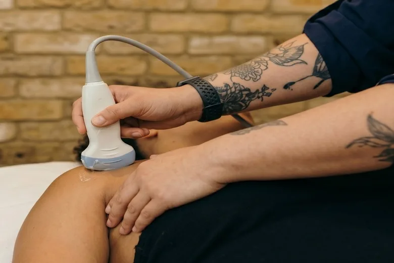
At Spine Plus Bow, we use advanced diagnostic musculoskeletal ultrasound to investigate injuries and conditions affecting muscles, tendons, ligaments, joints and surrounding soft tissues.
Ultrasound is a safe, non-invasive imaging technique that uses high-frequency sound waves to produce detailed, real-time images of structures beneath the skin. It allows us to identify a wide range of musculoskeletal abnormalities, including tendon tears, ligament injuries, joint inflammation, bursitis, muscle damage and degenerative changes.
The Advantage of Real-Time, Dynamic Imaging
One of the greatest advantages of musculoskeletal ultrasound is its ability to assess structures while they are moving.
Unlike conventional static imaging, such as MRI or X-ray, ultrasound allows us to examine tendons, muscles and joints dynamically. We can observe how tissues move, glide and interact during specific movements, often reproducing the patient's symptoms and helping to identify the underlying source of pain.
We can also compare the affected area directly with the opposite, unaffected side, helping us distinguish normal anatomical variations from genuine pathology.
Power Doppler imaging provides further information by identifying increased blood flow associated with certain inflammatory processes, such as active tendinopathy or synovitis.
Immediate Diagnosis and Treatment Planning
At Spine Plus Bow, diagnostic ultrasound is integrated directly into the clinical consultation.
Rather than waiting days or weeks for imaging appointments and reports, we can often perform an ultrasound examination during your initial appointment, explain the findings immediately and develop a personalised treatment plan.
Where appropriate, ultrasound can also be used to guide injections and other procedures, allowing treatment to be delivered accurately to the intended anatomical structure.
This streamlined approach can reduce unnecessary delays, support faster clinical decision-making and help patients begin appropriate treatment and rehabilitation sooner.
Common Conditions We Investigate
Musculoskeletal ultrasound can be particularly useful for assessing:
- Tendon injuries: Tendinopathy, partial and full-thickness tears, calcific deposits and tendon inflammation.
- Ligament injuries: Sprains, tears and ligament instability.
- Muscle injuries: Strains, tears, contusions and intramuscular haematomas.
- Joint conditions: Osteoarthritis, joint effusions, synovitis and capsulitis.
- Bursitis: Inflammation or fluid accumulation within bursae, including the shoulder, hip, knee and elbow.
- Nerve entrapments: Conditions such as carpal tunnel syndrome and selected peripheral nerve compressions.
- Soft tissue swellings: Ganglion cysts, Baker's cysts, lipomas and other superficial lumps.
- Inflammatory conditions: Assessment of synovial thickening, increased vascularity and soft tissue inflammation.
Ultrasound-Guided Injections and Procedures
In addition to diagnosis, ultrasound allows us to perform a range of precisely targeted treatments under direct visual guidance.
These may include:
- Corticosteroid injections for selected inflammatory joint, tendon sheath and bursal conditions.
- Hyaluronic acid injections for symptomatic osteoarthritis.
- Dextrose prolotherapy for selected musculoskeletal conditions.
- Ultrasound-guided tendon procedures and other targeted interventions where clinically appropriate.
By visualising the needle and surrounding anatomy in real time, we can improve placement accuracy while helping to avoid nearby nerves, blood vessels and other sensitive structures.
Safe, Convenient and Radiation-Free
Musculoskeletal ultrasound does not use ionising radiation, making it a safe and convenient imaging option for many patients.
The examination is generally comfortable, requires little or no preparation and can be repeated when clinically necessary to assess healing or monitor progress.
Although ultrasound is extremely effective for examining many superficial musculoskeletal structures, deeper structures, bone marrow, cartilage and certain spinal conditions may require MRI, CT or X-ray for further assessment.
Why Choose Spine Plus Bow
Our approach combines clinical examination, diagnostic ultrasound and targeted treatment within the same setting.
This means we can often identify the likely source of your symptoms, explain the findings and begin an appropriate management programme during a single visit.
Our aim is simple: to provide an accurate diagnosis, minimise unnecessary appointments and help you return to normal activity as quickly and safely as possible.